PHONE
+44-7482-878921
+44-7482-878921
2376-0249
Clinical-Medical Image - International Journal of Clinical & Medical Images (2023) Volume 10, Issue 6

Author(s): Bach-Bachich Lea*, Poropat Goran, Dora FuÄÂkar Čupić and Irena Krznarić Zrnić
1Department of Pediatrics, School of Medicine, University of Zagreb, Zagreb, Croatia
2Department of Gastroenterology and Hepatology, University Hospital Centre Rijeka, Croatia
3Department of Pathology and Cytology, University Hospital Center Rijeka, Croatia
*Corresponding Author:
Bach-Bachich Lea
Department of Pediatrics, School of Medicine
University of Zagreb, Zagreb, Croatia
E-mail: bachlea@gmail.com
Received: 23 May 2023, Manuscript No. ijcmi-23-99781; Editor assigned: 24 May 2023, Pre QC No. P-99781; Reviewed: 01 June 2023, QC No. Q-99781; Revised: 02 June 2023, Manuscript No. R-99781; Published: 09 June 2023, DOI:10.4172/2376-0249.1000899
Citation: Lea BB, Goran P, ÄÃâÃÅupiÄÃâÃâ¡ DF and ZrniÄÃâÃâ¡ IK. (2023) An Unusual Case of Gastric Heterotopia in the Ascending Colon. Int J Clin Med Imaging 10: 899.
Copyright: © 2023 Lea BB, et al. This is an open-access article distributed under the terms of the Creative Commons Attribution License, which permits unrestricted use, distribution, and reproduction in any medium, provided the original author and source are credited.
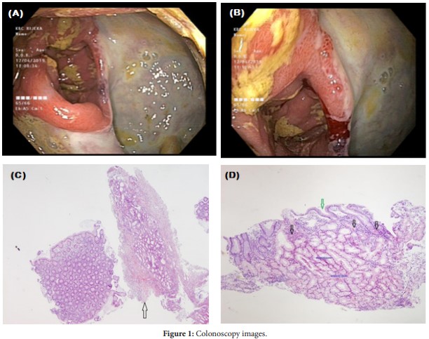
A 53-year-old male with a history of DBM type 2 and chronic pancreatitis was admitted through the ED for RLQ and epigastric pain with 20 days of blood-mixed diarrhea and no bowel movements in the past 3 days. The patient was severely malnourished (BMI 13), nonfebrile, did not travel recently and had no sick contacts. On physical exam bowel sounds were heard, there was diffuse abdominal tenderness without signs of peritonitis and negative LS. Parenteral/enteral nutrition was administered. Microbial stool analysis was negative. CT scan showed mild thickening of the gastric and small bowel wall, significant calcification of pancreatic tissue due to chronic pancreatitis and notable atherosclerosis of the aorta and its visceral branches. A large ulcerative lesion covering more than 50% of the circumference of the ascending colon was found on colonoscopy (Figure 1A,B). On microscopic examination of the biopsies, six samples were consistent with colonic mucosa while two were found to contain gastric mucosa with shortened pits lined by specialized foveolar epithelium (green arrow) and glands lined by mainly mucinous (blue arrows) and red parietal cells (black arrows) (Figure 1C,D).
Gastric heterotopia; Endoscopy; Colonoscopy; Colonic ulcer; Gastric heterotopia in the ascending colon; Biopsy; Histology; Ascending colon; Colonic pathology; Ulcerative lesion; Gastric mucosa; Parietal cells
None of the authors has any conflicts of interests to disclose.
 Awards Nomination