international journal of clinical & medical images, clinical journals, medical journals, omics international, international journals, open access publication, scientific journals, free online medical journals, peer reviewed online journals, research, free online scientific articles
PHONE
+44-7482-878921

International Journal of Clinical & Medical Images

2376-0249

Case Blog - International Journal of Clinical & Medical Images (2016) Volume 3, Issue 10

Band-like Acral Osteolysis in Limited Cutaneous Scleroderma

Band-like Acral Osteolysis in Limited Cutaneous Scleroderma

Author(s): Synrang B Warjri, Baphiralyne Wankhar, Animesh Mishra, Bhupen Barman, Tony Ete,

Abstract: Acral osteolysis is a well-recognized manifestation of scleroderma. Scleroderma is characterized by abnormal deposition of collagen and other extra cellular matrix macromolecules leading to vascular damage, inflammation and tissue fibrosis. Acral osteolysis in scleroderma occurs due to digital ischemia resulting from the vasculopathy. Treatment is directed towards providing symptomatic relief and prevention of complications. Here we present a case of band-like acral osteolysis seen in a patient of limited cutaneous scleroderma.

Keywords: Osteolysis; Acral osteolysis; Scleroderma; Systemic sclerosis

Introduction:Scleroderma is a multisystem connective tissue disorder that is characterised by abnormal deposition of collagen and other connective tissue macromolecules in the skin and multiple internal organs leading to inflammation and fibrosis. The exact etiology is unknown. Various genetic, infectious, and environmental factors have been implicated in the pathogenesis of scleroderma leading to vascular injury, fibrosis, and immune activation. The pathological pathways include vasculopathy and abnormalities in both humoral and cellular immunity. Osteolysis is a well-recognized manifestation of scleroderma. Here we report a case of band-like acral osteolysis seen in a patient of scleroderma.

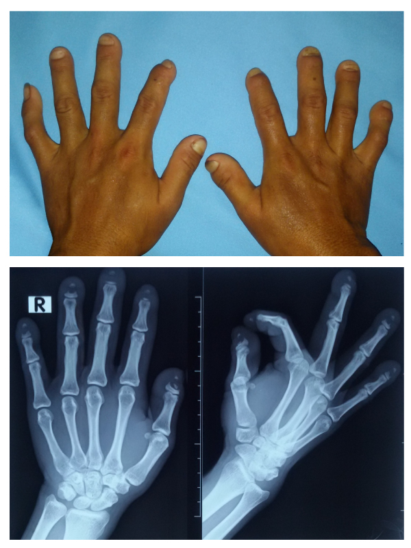
Clinical Presentation: A 25 years old female presented with a history of progressive tightening of the skin over her fingers and face for 3 years associated with phasic changes in skin colour (white, blue and red) typical of Raynaud’s phenomenon. On physical examination, the skin over her face appear stretched out, tight and shiny. The skin over the distal part of most her fingers also appear tight and stretched out. The tips of most of her fingers appear shortened (Figure 1). A test for antinuclear antibodies was positive with a titre of 1: 1280. Her Anticentromere antibodies were positive. A radiograph of both hands showed symmetrical band-like osteolysis of the diaphysis of distal phalanges suggestive of acral osteolysis (Figure 2). Echocardiography revealed a normal study. Further test were done that ruled out other organ systems involvement.

Discussion: Acral osteolysis in scleroderma is thought to result from impaired blood flow due to vasculopathy resulting in digital ischemia and subsequent damage to the terminal phalanges [1]. It may also be a result of pressure changes due to the induration and tightening of the skin. Acral osteolysis can also be seen in various other pathological conditions such as diabetes mellitus, tabes dorsalis, leprosy, psoriasis, rheumatoid arthritis, exposure to vinyl chloride, following trauma or in association with hyperparathyroidism [2].
Treatment options are limited and is focused on providing symptomatic relief and preventing complications. Patients should avoid prolonged cold exposure and avoid digital or skin trauma. Calcium channel blockers and alpha receptor blockers may reduce the ischemia by promoting vasodilatation. Phosphodiesterase-5 inhibitors such as sildenafil may also help provide symptomatic relieve. The disease is, however, progressive in nature and currently there are no approved therapy aimed at retarding the vascular damage, fibrosis and altered immunological response. Current experimental therapies are based on immune-suppression and immune-modulation and include drugs like methotrexate, mycophenolate mofetil, D-penicillamine and rituximab. Tocilizumab, an interleukin-6 (IL-6) receptor antagonist was recently granted breakthrough therapy designation status for systemic sclerosis by the United States Food and Drug Administration (USFDA) [3]. Various other possible immune-inflammatory therapies are currently under study, including modulators of the profibrotic pathways [4]. The safety and possible toxicity of these agents remain a cause for concern.


flyer Image Awards Nomination
Indexing and Archiving
A generic square placeholder image with rounded corners in a figure.
All published articles are assigned to Digital Object Identifier (DOI)- CrossRef.
A generic square placeholder image with rounded corners in a figure.
All published articles of this journal are included in the indexing and abstracting coverage of:
Google Scholar citation report
Citations : 293

International Journal of Clinical & Medical Images received 293 citations as per Google Scholar report