2376-0249
Medical Image - International Journal of Clinical & Medical Images (2015) Volume 2, Issue 3

Author(s): Alper Kaya* and Beyza Kaya
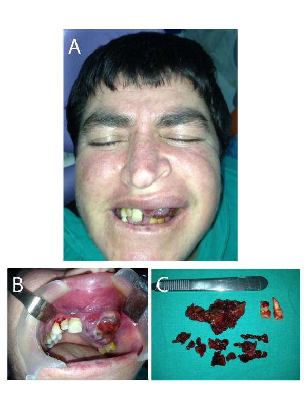
Medical Image: A 36 year old female patient was referred to our clinic with a chief complaint of swelling in the anterior maxilla which has been noticed seven months ago and rapidly attained the present size (Figure 1). According to the patient’s relatives’ report (because of the patient was mental retarded); this growth had appeared after extraction of tooth 21. The patient was taken to Ear-Nose-Throat clinic about three months ago. After a biopsy she was not treated surgically, only an antibiotic had been prescribed. The growth had not been regressed, contrarily it continued enlarge. Intraoral clinical examination showed a swelling extending from teeth 11 to 25 obliterating the buccal sulcus, measuring 6x4x3 cm with a firm and erythematous surface (Figure 2). The Computerized Tomography scan revealed an unilocular radiolucency extending to basis of orbita and nose. Surgery was performed under local anesthesia (Figure 3). The tissue was removed and the histopathological diagnosis was a CGCG. Histopathologic examination reveals ulcerative paraceratotic connective tissue with a proliferation of osteoclast-type multinucleated giant cells and granulation tissue rich in mononuclear inflammatory cells and hemosiderin pigments. Unfortunately, we are unable to report after surgery because contact with the patient has been lost.
 Awards Nomination

