2376-0249
Powerpoint Presentation - International Journal of Clinical & Medical Images (2015) Volume 2, Issue 3

Author(s): Massimo Bolognesi*
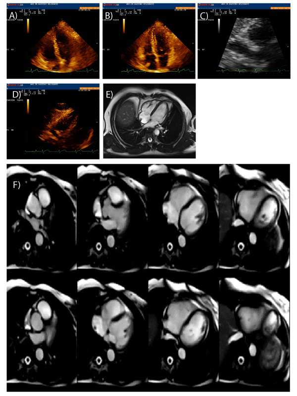
The differential diagnosis of intracardiac masses that involves the right atrial chamber include vegetation, thrombus or tumours, and anatomic normal variants such as crista terminalis. Size, shape,location, mobility and attachment of the mass combined with the clinical findings help differentiate etiology. Two Dimensional Trans Thoracic Echocardiography (TTE) became the gold standard test for the diagnosis of intracardiac masses and further examination such as transesophagealechocardiography (TEE) and Cardiac Magnetic Resonance (CMR) improved the accuracy. The cardiovascular imaging is crucial to establish a correct diagnosis for proper management and therapy. Here the author describes the case report of a healthy middle-aged athlete who underwent echocardiography for sports preparticipation screening which detected the presence of a peduncolated right atrial mass of uncertain nature. Because of the strong suspicion for small right atrial mixoma, he underwent CMR that showed a small enhancing mass adherent to the right atrial posterior wall typical of prominent crista terminalis. This report of rare Crista Terminalis muscular bridge that mimicks right atrial mixoma gives new information for the understanding of the aspect of complex and strange right atrial anatomy
 Awards Nomination

