PHONE
+44-7482-878921
+44-7482-878921
2376-0249
Clinical Image - International Journal of Clinical & Medical Images (2021) Volume 8, Issue 2

Author(s): Olaia Chalh*, Safae Choayb, Nazik Allali, Latifa Chat and Siham Elhaddad
Clinical Image
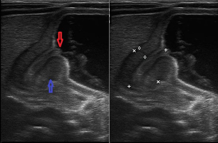
Hypertrophic pyloric stenosis is the most common surgical cause of postprandial vomiting in early infancy. It has an incidence of 3/1000 live births per year and occurs mostly in males with a range age from 2 to 6 weeks of life. This condition is defined by thickening of the muscular layer and failure in relaxation of the pyloric canal. The diagnosis can be made clinically by the presence of palpable pyloric ‘olive’ in the right upper quadrant of the abdomen. When physical findings alone are inconclusive, abdominal ultrasound should be performed. It shows a thickened is the most precise diameter. On longitudinal views, the pylorus had an appearance of uterine cervix and described as a ‘‘cervix sign’’. Prominent hyperechogenic mucosa projecting into the antrum is known as the ‘‘antral nipple sign’’ (Figure 1).
Keywords: Pyloric stenosis; Ultrasound
 Awards Nomination