PHONE
+44-7482-878921
+44-7482-878921
2376-0249
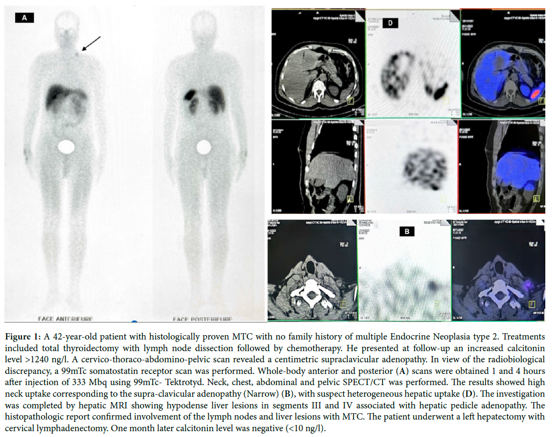
Clinical Image - International Journal of Clinical & Medical Images (2024) Volume 11, Issue 5

Author(s): Amellouk Sara*, Bennani Hakim, Bensmimou Hafsa, Batani Halima, Oussafrar Zakaria and Guensi Amal
Department of Nuclear Medicine, University Hospital Center Ibn Rochd, Casablanca, Morocco
Received: 01 May 2024, Manuscript No. ijcmi-24-133937; Editor assigned: 03 May 2024, Pre QC No. P-133937; Reviewed: 17 May 2024, QC No. Q- 133937; Revised: 23 May 2024, Manuscript No. R- 133937; Published: 31 May 2024, DOI:10.4172/2376-0249.1000956
Citation: Sara A, Hakim B, Hafsa B, Halima B, Zakaria O, et al. (2024) Comparison of Octreotide Scintigraphy and Conventional Imaging in Detection of Metastatic Medullary Thyroid Carcinoma Case Report. Int J Clin Med Imaging 11: 956.
Copyright: © 2024 Sara A, et al. This is an open-access article distributed under the terms of the Creative Commons Attribution License, which permits unrestricted use, distribution, and reproduction in any medium, provided the original author and source are credited.
Medullary Thyroid Carcinoma (MTC) is a rare neuroendocrine tumor arising from the parafollicular C cells of thyroid. It represents approximately 5% of all thyroid carcinomas. MTC is frequently aggressive and metastatic sites are generally cervical and mediastinal lymph nodes, lungs, liver, and bones. The initial treatment consists of a total thyroidectomy with bilateral neck lymph node and upper mediastinum dissection [1]. Somatostatin receptors are often over-expressed in Medullary Thyroid Carcinoma (MTC), the aim of our study was to evaluate the utility of scintigraphy with the somatostatin analogue 99mTc- Tektrotyd in metastatic MTC in comparison with other conventional imaging techniques. Nuclear imaging modalities are sensitive in detecting small amounts of residual or recurrent tumor at an early stage that may not be visible on conventional imaging, helping to guide treatment decisions and improve patient outcomes [2]. Normalization of serum calcitonin levels after surgery is a strong indicator that neoplastic tissue was totally removed.
Metastatic medullary thyroid carcinoma, Octreotide scintigraphy, conventional imaging
The authors certify that they have obtained all appropriate patient consent forms. In the form, the legal patient has given his consent for images and other clinical information to be reported in the journal.
Nil.
There are no conflicts of interest.
[1] Wahl RA, and Röher HD. (1988). Surgery of C cell carcinoma of the thyroid. In Thyroid tumors Vol. 19: 100-112.
Google Scholar, Crossref, Indexed at
[2] Skoura E. Depicting medullary thyroid cancer recurrence: The past and the future of nuclear medicine imaging. Int J Endocrinol Metab, (2013): 11(4).
 Awards Nomination