PHONE
+44-7482-878921
+44-7482-878921
2376-0249
Clinical Image - International Journal of Clinical & Medical Images (2014) Volume 1, Issue 4

Author(s): Int J Clin Med Imaging 2014
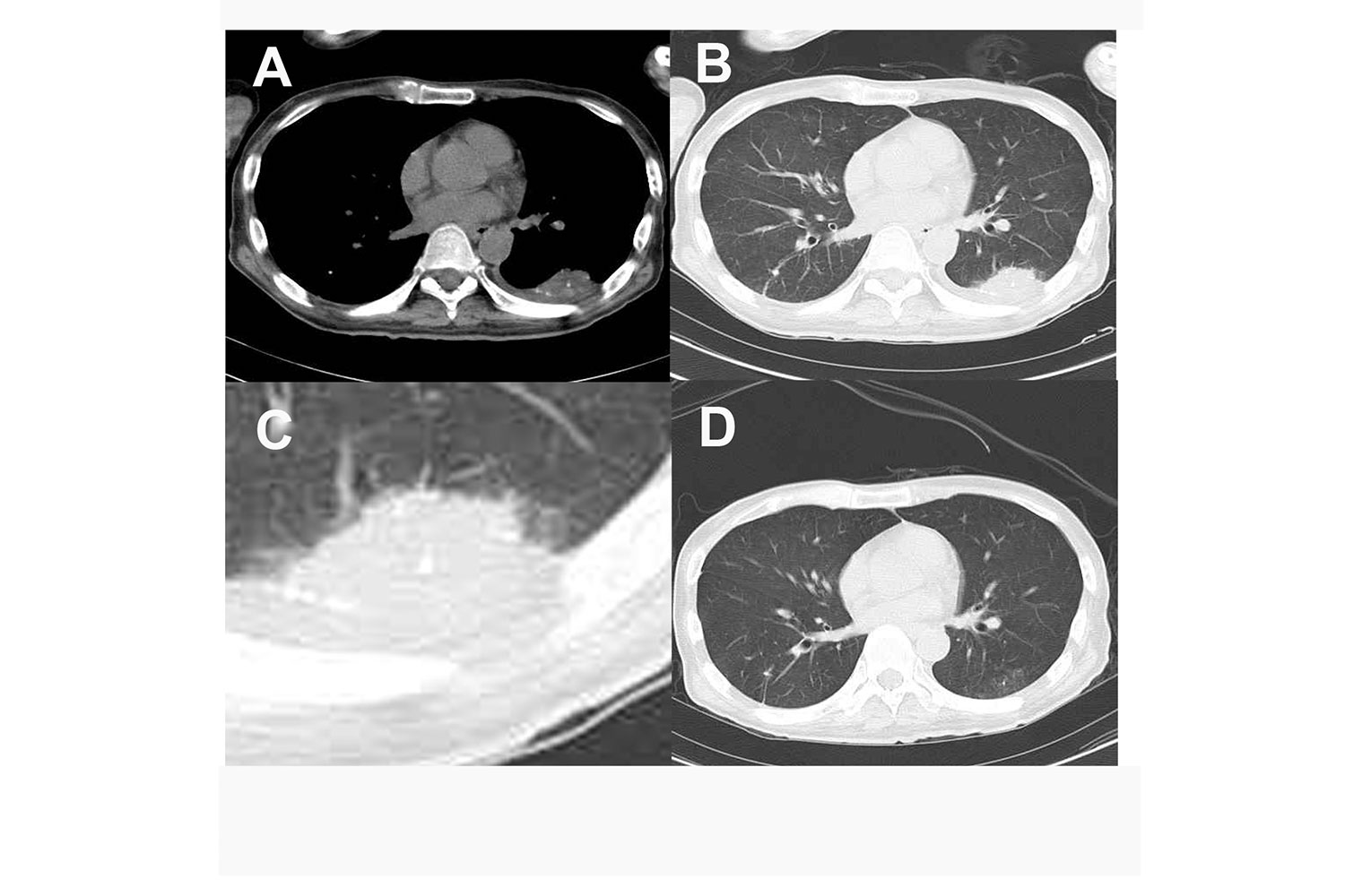
A 44-year-old Japanese man with chorea acanthocytosis underwent a video fluorographic study of swallowing (VF) in which he was required to Nonionic Soluble Contrast Media (NSC). After examination, all his nutritional requirements were met by tube feeding and not by oral feeding. One month after VF, he underwent a Computed Tomography (CT) examination of the chest because of increased sputum without fever. A CT scan showed a consolidation including higher density dots similar to calcification in the left lower lobe, indicating pneumonia by the aspiration of NSC (Figure A-C). In general, when NSC is used for bronchoscopy, complete clearance of NSC from the lungs usually occurs within 24 h [1,2]. The sputum samples tested negative for tuberculosis on laboratory examination. The consolidation disappeared after 1 month without any medication (Figure D).
 Awards Nomination