PHONE
+44-7482-878921
+44-7482-878921
2376-0249
Clinical-Medical Image - International Journal of Clinical & Medical Images (2022) Volume 9, Issue 8

Author(s): Belkouchi Lina*, Iraqi Houssaini Zaynab, Khamlichi Amina, Fikri Meriem, El Kettani Najwa, Jiddane Mohamed and Touarsa Firdaous
Department of Neuroradiology, Hôpital des spécialités de Rabat, Faculty of Medicine and Pharmacy of Rabat, Mohammed V University Hospital, Rabat, Morocco
Date of Submission: 15 August 2022, Manuscript No. ijcmi-22-74848; Editor assigned: 16 August 2022, Pre QC No. P-74848; Reviewed: 22 August 2022, QC No. Q-74848; Revised: 25 August 2022, Manuscript No. R-74848; Published: 31 August 2022, DOI: 10.4172/2376-0249.1000845.
Citation: Lina B, Zaynab EH, Amina K, Meriem F, Najwa EK, et al. (2022) Dyke Davidoff Masson Syndrome: Typical Aspect. Int J Clin Med Imaging 9:845.
Copyright: © 2022 Lina B, et al. This is an open-access article distributed under the terms of the Creative Commons Attribution License, which permits unrestricted use, distribution, and reproduction in any medium, provided the original author and source are credited.
Dyke Davidoff Masson syndrome (DDMS), is a rare neurological condition first described in 1933 by Dyke, Davidoff and Masson after evaluating nine patients presenting the following clinical symptoms: Facial asymmetry, hemiparesis, seizures and intellectual disability with unusual skull radiographs and pneumatoencephalographic changes [1-3].
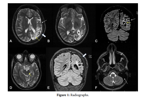
Its pathogenesis remains unclear, however some authors suggested its link to in utero or post natal brain injuries due to hemorrhage, trauma, infection, or vascular lesions. Clinical symptoms include mainly: Facial asymmetry, hemiparesis, hemiplegia, seizures, cognitive and psychiatric symptoms, and they all depend on the extent of the lesions. Seizures may occur years after hemiparesis and they can be focal or generalized. Imaging is the examination of choice to confirm diagnosis and evaluate severity. Modalities include either a CT or an MRI. Typical aspects include: Cerebral hemi atrophy with dilation of ipsilateral lateral ventricle with compensatory calvarial thickening (thickening of the adjacent skull bone) and sinuses enlargement especially the frontal sinus and mastoid air cells (Figure 1).
The images above represent a 25 years old male patient, admitted for generalized seizures associated with a right sided hemiparesis, the MRI showed:
• Left hemispheric atrophy (White arrow, Figure 1A and 1C).
• Focal encephalomalacia of the left parieto-occipital lobe associated to gliosis (Yellow arrow, Figure 1A and 1C).
• Ipsilateral dilation and attraction of lateral ventricle (Star, Figure 1B and 1C).
• Reduced volume of the left cerebral peduncle (Yellow arrow, Figure 1D).
• Compensatory calvarial thickening on the left side (Thick white arrows Figure 1A and 1E).
• Enlargement of frontal sinus and mastoid air cells compared to the right side (Arrows, Figure 1F).
Hemiatrophy; Cerebral; Neurology; Imaging
[1] Jain MJ and Kovela RK. (2021) Dyke-Davidoff-Masson syndrome. Pan Afr Med J 39.
[2] Al-Smair A, Hafez SA, Saadeh A and Al-Ali A. (2022) An adult with Dyke–Davidoff–Masson Syndrome: A case report. Cureus 14(3).
[3] Bhol D, Chandrasekar S, John J and Satapathy A. (2021) Dyke–davidoff–masson syndrome: A rare cause of acquired cerebral hemiatrophy. Asian J Neurosurg 16: 579-581.
 Awards Nomination