PHONE
+44-7482-878921
+44-7482-878921
2376-0249
Clinical-Medical Image - International Journal of Clinical & Medical Images (2022) Volume 9, Issue 8

Author(s): Lemrabet Abir*, Bahlouli Nourrelhouda, L Jeroundi and FZ Laamrani
Department of Emergency Radiology, CHU Ibn Sina, Rabat, Morocco
Date of Submission: 24 August 2022, Manuscript No. ijcmi-22-72772; Editor assigned: 25 August 2022, Pre QC No. P-72772; Reviewed: 26 August 2022, QC No. Q-72772; Revised: 27 August 2022, Manuscript No. R-72772; Published: 31 August 2022, DOI: 10.4172/2376-0249.1000842
Citation: Abir L, Nourrelhouda B, Jeroundi L and Laamrani FZ. (2022) Gamna-Gandy Bodies: A Case Report. Int J Clin Med Imaging 9:842.
Copyright: © 2022 Abir L, et al. This is an open-access article distributed under the terms of the Creative Commons Attribution License, which permits unrestricted use, distribution, and reproduction in any medium, provided the original author and source are credited.
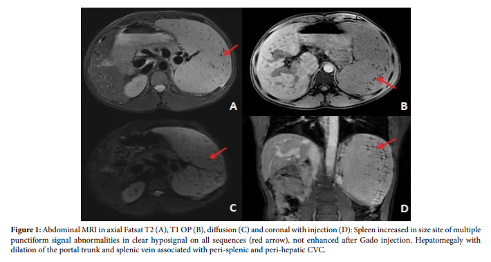
A 33-year-old patient, followed for chronic liver disease with signs of portal hypertension and in whom the biological assessment found pancytopenia with negative hepatic serology. Faced with the sudden appearance of hepatic colic, an abdominal ultrasound was performed. Carried out in emergency objectifying a dilation of the intra and extra hepatic bile ducts without sonographically detectable obstacle. An additional MRI with injection of PDC was requested in search of an etiology had shown no biliary obstacle. Splenic nodular signal abnormalities were highlighted on this MRI in connection with gamma corpuscles Gandy (Figure 1).
Gamna–Gandy bodies (GGBs) are fibrosiderotic nodules presenting as small necrotic-hemorrhagic areas in the spleen most commonly caused by portal hypertension. However, they can be seen in other conditions such as sickle cell anemia, paroxysmal nocturnal hemoglobinuria and splenic vein thrombosis.
stonemarketConsequences of a micro haemorrhage occurring after sinus rupture in a congestive parenchyma leading to a deposition of hemosiderin and calcium followed by a fibroblastic reaction. Ultrasound shows hyperechoic punctiform lesions disseminated throughout the splenic parenchyma with or without shadow cone. CT scan identifies strong attenuation foci that are indistinguishable from splenic granulomas. MRI is the most sensitive technique for detecting these nodules because of their iron content. They are typically hypo signal on all sequences with no enhancement. GGBs have no specificity in cirrhosis or portal hypertension [1-4].
Gamna-Gandy bodies; Portal hypertension; MRI
[1] Selcuk D, Demirel K, Kantarci F et.al. (2005) Gamna-Gandy bodies: a sign of portal hypertension. Turk J Gastroenterol 16: 150-152.
Google Scholar, Crossref, Indexed at
[2] Luna A, Ribes R, Caro P et.al. (2006) MRI of focal splenic lesions without and with dynamic gadolinium enhancement. American J R 186: 1533-1547.
Google Scholar, Crossref, Indexed at
[3] Chan YL, Yang WT, Sung JJ, Lee et.al. (2000) Diagnostic accuracy of abdominal ultrasonography compared to magnetic resonance imaging in siderosis of the spleen. J Ultrasound Med, 19: 543-547.
Google Scholar, Crossref, Indexed at
[4] Luo TY, Itai Y, Yamaguchi M et.al. (1998) Gamna-gandy bodies of the spleen depicted by unenhanced CT: report of two cases. Radiat Med 16: 473-476.
 Awards Nomination