PHONE
+44-7482-878921
+44-7482-878921
2376-0249
Clinical-Medical Image - International Journal of Clinical & Medical Images (2021) Volume 8, Issue 6

Author(s): Belkouchi Lina, Bourekba Iliass, Outznit Mustapha, Nazik Allali, Latifa Chat, El-Haddad Siham
Clinical Image
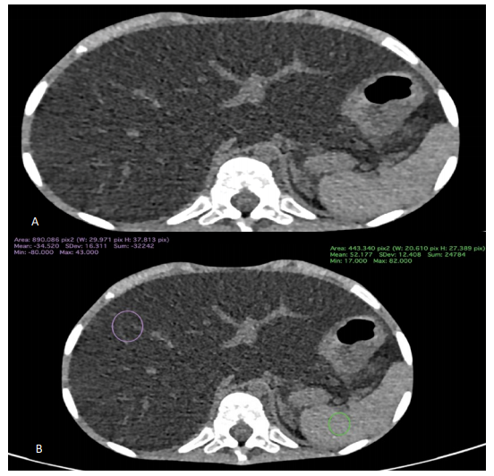
Hepatic steatosis is the presence of fat vacuoles in hepatocytes. Liver biopsy was used to confirm diagnosis, however being invasive with complication risks, imaging have become a non-invasive method of choice to confirm diagnosis and even quantify hepatic steatosis, allowing the evaluation of the liver parenchyma in a wider range. Imaging modalities include ultrasonography (US), CT-scan and MRI. US may lead to diagnosis if over 20% of liver parenchyma contains fat vacuoles. This will be revealed through the presence of hepatomegaly associated to an increased echogenicity of the liver compared to the renal cortex. MRI is considered as the examination of choice to diagnose and quantify liver fat; however, it is not always available. CT-scan is an alternative imaging method, being more accessible with fast acquisitions. It has been considered as a non-invasive reference method, along with the MRI, in the diagnosis of hepatic steatosis. The accumulation of intra cellular fat is revealed in unenhanced CT as a reduced attenuation of liver parenchyma compared to the spleen. Liver parenchyma attenuation value normally exceeds the spleen’s by 8-10 HU, in unenhanced CT. Measurement is done by manually placing a ROI (Region Of Interest) in the right hepatic parenchyma and the spleen parenchyma (Figures 1A and 1B); A ratio 30% of liver parenchyma fat. Enhanced CT scan has proven its efficiency in quantifying hepatic steatosis between mild moderate or severe, and is based on liver contrast attenuation, with or without considering splenic attenuation. In contrasted CT at a portal phase between 80-100s the presence of a difference of attenuation between liver parenchyma and the spleen> 20HU is almost specific of hepatic steatosis. CT-scan is nowadays a reference standard to detect moderate to advanced liver steatosis.
Keywords: Liver; Steatosis; Imaging; CT
 Awards Nomination