2376-0249
Case Blog - International Journal of Clinical & Medical Images (2016) Volume 3, Issue 6

Author(s): Karthik Shunmugavelu
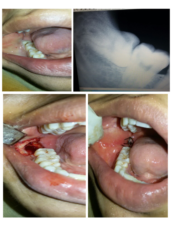
Clinical Presentation: A 35 year old female presented to the Department of Dentistry and Faciomaxillary Surgery complaining of discomfort in the right mandibular third molar region (fourth quadrant). General health examination revealed that the patient was conscious, oriented, afebrile and vitals were stable. On intraoral clinical examination, partially clinically exposed right mandibular third molar was present along with pericoronitis, halitosis, pain, swelling and gingival bleeding (Figure 1). Patient stated that the discomfort has been present since a week. The pain increased in intensity during mastication. Radiographically, horizontaly impacted right mandibular third molar was seen in radiovisiogram (RVG) (Figure 2). Treatment procedure included planned surgical removal of the specific tooth under local anaesthesia with 1:2,00,000 adrenaline. After placement of mouth prop, Fraser suction in place, topical local anesthetic applied, inferior alveolar nerve block was administered , incision placed with the no.3 and no.11 Bard Parker surgical handle and blade, mucoperiosteal flap raised with the help of periosteal elevator, overlying bone and the impacted tooth clinically seen (Figure 3). The flap was retracted with the help of Austins retractor whereas the tongue by tongue depressor. With the help of micromotor handpiece apparatus which includes, high speed sharp carbide burs, no.557/703 fissure burs, no.8 round bur, excess bone coverage was trimmed so that the horizontally placed tooth was surgically uplifted by wedging with the help of Couplands elevator in the fulcrum region which lies between distal surface of right mandibular second molar and the mesial coronal surface of the impacted tooth. To reduce the heat production during bone cutting, 18 gauge needle and 10 ml syringe were used for saline irrigation. After removal of the impacted tooth in full portion with the help of lower molar forceps (Figure 4), the socket was curetted for fresh bleeding (Figure 5), bony margin were trimmed by bone rongeur, bone smoothening done by bone file, mucoperiosteal flap replaced, soft tissue held by Adson forceps and 3-0 black silk sutures placed (Figure 6) with the help of needle holder along with cutting edge suture needle and excess suture material was removed by Metzenbaum scissors. After a week of review, the sutures were removed with an evidence of excellent intraoral healing.
 Awards Nomination

