PHONE
+44-7482-878921
+44-7482-878921
2376-0249
Clinical Image - International Journal of Clinical & Medical Images (2021) Volume 8, Issue 1

Author(s): Lina Belkouchi*, Kaoutar Imrani, Mustapha Outznit, Hounayda Jerguigue, Rachida Latib, and Youssef Omor
Clinical Image
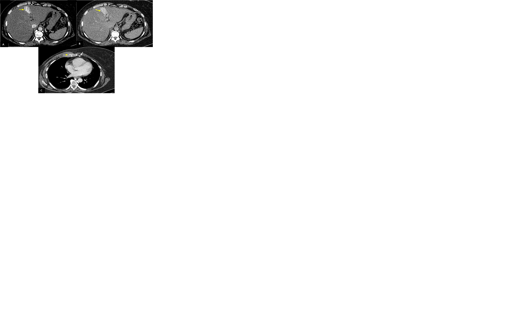
The hot spot sign is a radiological sign found in a contrasted CT-scan or technetium 99 m sulfur colloid scans. It was first described in 1983 by Ishikawa. It corresponds to a focal well shaped enhanced area in arterial or portal phase within the quadrate lobe of the liver also known as the IVb segment of the Bismuth-Couinaud classification system. It was first diagnosed in a superior vena cava syndrome and is caused by portosystemic venous shunt between the superior vena cava (SVC) and portal vein, mostly due to the obstruction of the SVC. Thus, it reflects an increased blood flow due to the shunting. It is an indicator of a thoracic central venous obstruction, as seen in the images above, showing a hot spot sign caused by the right internal mammal vein obstruction secondary to the recurrence of a breast cancer invading the internal mammal vessels (Figure 1).
Keywords: Hot spot sign; Quadrate lobe liver thoracic; Venous obstruction imaging
 Awards Nomination