PHONE
+44-7482-878921
+44-7482-878921
2376-0249
Case Blog - International Journal of Clinical & Medical Images (2015) Volume 2, Issue 7

Author(s): Maryam Poursadeghfard M*, Kazemi KH and Karamimagham S
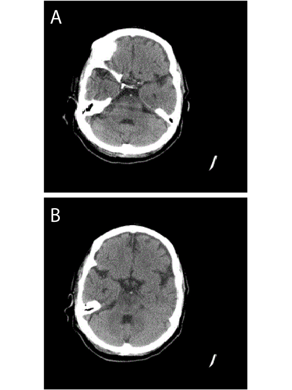
Posterior circulation is a vital area of brain blood supply and stroke in this territory could be fatal or result in severe disability. Many patients have preceding symptoms (vertigo or head ache) 2 weeks before [1]. Although brain CT scan is a rapid standard method to detect infarction after the first presentations, because of bony structures and artifacts, it is less sensitive for the posterior fossa and brain stem parenchyma. As brain MRI or brain vessels imaging (CTA, MRA) are not easily available tools for patients’ evaluation, fine informative changes on brain CT scan contributes to physicians’ early and more effective intervention. Hyperdense basilar artery sign is a sign of basilar artery thrombosis or embolism that predicts basilar territory infarction [2]. Here, we describe a 70 year-old lady who was referred to the hospital with sudden impaired level of consciousness. One day before, she had developed the right upper limb weakness and slurred speech and in the previous week she had new onset of headache and vertigo. Past medical history was consistent with ischemic heart disease and previous ischemic stroke 2 years ago. Brain CT showed hyper-dense signal change in the basilar artery indicative of basilar artery thrombosis and pons infarction (Figure 1a and 1b).
Impaired blood supply of the basilar artery causes devastating results and by the time the stroke develops, it is too late to approach appropriately. Non- enhanced brain CT scan is a very useful tool for early evaluation of patients with acute infarction; however, in the brain stem area and for basilar artery territory, it has some limitations because of bony artifacts in the skull base and most of the time further modalities are needed to document brain stem ischemia. So rapid and correct diagnosis just based on non-enhanced CT scan is sometimes a challenge especially for inexperienced physician.
 Awards Nomination