PHONE
+44-7482-878921
+44-7482-878921
2376-0249
Clinical Image - International Journal of Clinical & Medical Images (2017) Volume 4, Issue 4

Author(s): Mohammad S and HosseiniB M
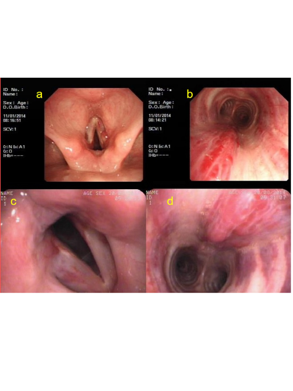
Clinical Image: A 23-year-old non-smoker woman presented with a seven-year history of intermittent hemoptysis, which always subsided spontaneously. There were no other symptoms and sources of bleeding. Her past medical, drug and familial history were unremarkable. Physical examination revealed nothing abnormal. Results from Para-clinical tests (CBC, U/A, S/E, Pulseoximetry, ABG, CXR, Air Bubble Contrast Echocardiography and pulmonary arterial pressure) were normal except showing a normocytic anemia. Laryngoscopy showed a nodule (five mm in diameter) and bloody strains on vocal cord (Figure 1A). Fiber-optic bronchoscopy showed telangiectatic lesions in the subglottic area to two-third of the proximal trachea (Figure 1B). The lesions were flat, non-pulsatile, and without active bleeding, surrounded by normal mucosa. One-third of distal trachea and main and lobar bronchi were normal. We diagnosed as Isolated Tracheal Telangiectasia. We performed palliative therapy with the Argon plasma coagulation device in three sessions (at first, second and eighth weeks). At a Six-month follow-up, there was no hemoptysis anymore and no problem in speech; Bronchoscopy showed vocal cord and tracheal lesions were less severe (Figure 1C and D). The patient remains asymptomatic in a one-year follow-up.
 Awards Nomination