2376-0249
Clinical Image - International Journal of Clinical & Medical Images (2016) Volume 3, Issue 6

Author(s): Deepak S, Warrier K, Kerslake R and Rangaraj S
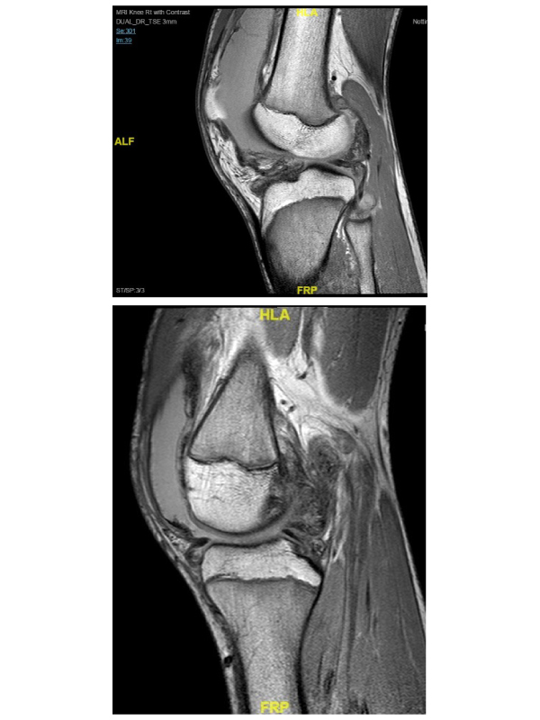
A 14 year old previously fit and well boy was referred by the orthopaedic team with a 7 month history of swelling in right knee. Aspiration of the knee joint revealed blood stained fluid, which was negative for bacterial cultures. The swelling and pain progressively got worse, with no systemic symptoms including fever. Further review by the orthopaedic team was followed by blood tests, which showed normal inflammatory markers. MRI of his right knee was reported to show a multi-loculated fluid collection with septae and post contrast enhancement of the collection walls but no involvement of underlying bone. He also had a synovial biopsy which showed synovial thickening and fibroid degeneration and no evidence of granuloma and malignancy. He was then referred to paediatric rheumatology team to consider Juvenile Idiopathic Arthritis (JIA). On examination he had a large swelling of the right knee joint more prominent in his supra patellar region with warmth and minimal restriction of movement. The rest of the joints were normal. In view of the history and clinical course of a teenage boy with large effusion affecting a single knee joint with blood stained synovial fluid, a diagnosis of pigmented villonodular synovitis (PVNS) was considered. He had a repeat MRI, which reported prominent and in some places nodular low signal intensity synovial proliferation around the knee and a large effusion, consistent with PVNS. He was referred back to the orthopaedic team after poor response to intraarticular steroid injection and is currently awaiting synovectomy.
Pigmented villonodular synovitis (PVNS) is a benign proliferative disorder of uncertain etiology that affects synovial lined joints, bursae and tendon sheaths (Figures 1 and 2), most commonly in the 20-50 years age group, without gender predilection [1]. The disorder results in various degrees of villous and/or nodular changes in the affected structures. PVNS lesions are monoarticular or solitary. Polyarticular disease is uncommon condition. Although a benign condition, PVNS may result in significant morbidity if left untreated. Pain, loss of function, and eventual joint destruction may result. The primary treatment options include surgical resection via synovectomy or radiation therapy [2-3]. The MRI findings
 Awards Nomination

