PHONE
+44-7482-878921
+44-7482-878921
2376-0249
Clinical Image - International Journal of Clinical & Medical Images (2016) Volume 3, Issue 5

Author(s): Mukund Das, Chirag Rana, Salvatore Zisa, Fayez Shamoon and Nagwa Hafez
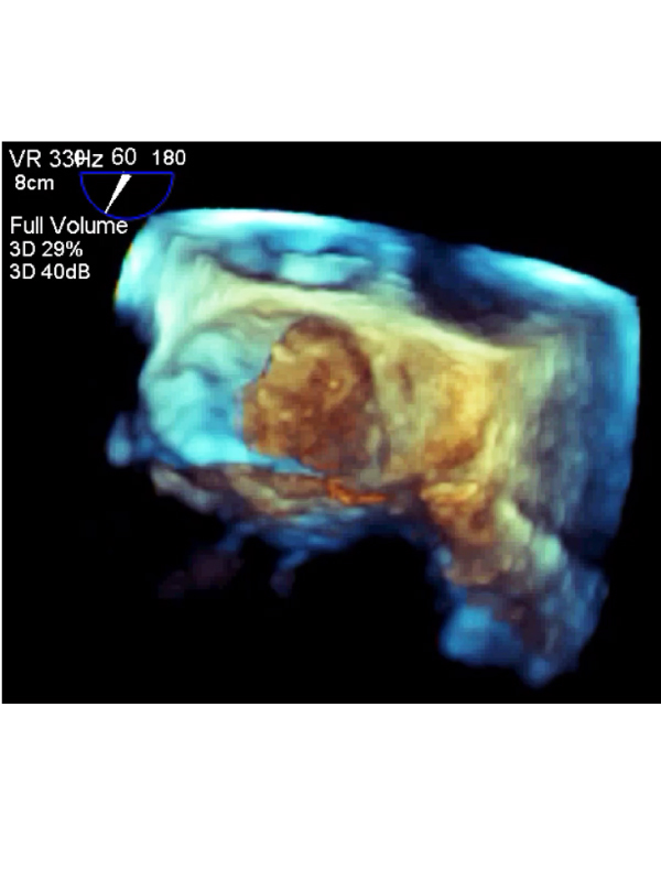
A 74-year-old female presented with her second episode of stroke over the past two weeks. During the work-up, a left atrial myxoma was discovered and was found to be the cause of embolic phenomenon that caused her previous strokes. The patient underwent surgical excision of the tumor. Here, we are showing (Figure) the myxoma on 3D-transesophageal echocardiography (TEE) that was obtained intra-operatively. The myxoma can be seen attached to the interarterial septum. The mass is irregularappearing, non-pedunculated, and not causing any mitral valve (black arrow) obstruction. Note the area of loose-flapping tumor fragment at the 11’O clock position of the tumor (white arrow). It is likely that such loose-flapping tumor fragments were embolizing to her cerebral vessels and presenting as stroke. Myxomas are the most common type of benign cardiac tumors. Although they are benign in nature, they may causes catastrophic events from tumor embolization, mitral valve obstruction, or myocardium invasion.
 Awards Nomination