PHONE
+44-7482-878921
+44-7482-878921
2376-0249
Case Blog - International Journal of Clinical & Medical Images (2016) Volume 3, Issue 3

Author(s): Donboklang Lynser
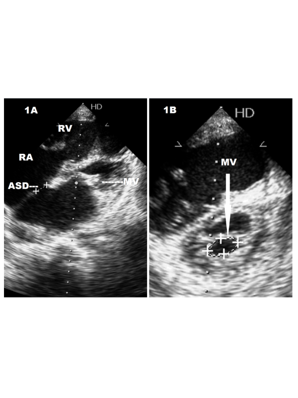
Figure 1: Echocardiography in a 40 year old female of lutembachers syndrome. A. Showing the atrial septal defect (ASD) with mitral valve thickening and doming. B. Cross-section of the mitral valve (MV) showing mitral stenosis.
Figure 2: Echocardiography with colour flow imaging in lutembacher’s syndrome. A. Colour Doppler flow imaging showing flow across the ASD from left to right atrium. B. Showing pulmonary regurgitation (PR) jet.
Abstract Lutembacher’s syndrome is defined as congenital atrial septal defect with acquired mitral stenosis. We present echocardiographic findings in a 40 year old female of lutembacher’s syndrome.
Clinical Presentation A 40 year old female patient presented with on and off episodes of breathlessness. Clinically there is evidence of raised Jugular venous pressure (JVP), peripheral pitting edema and tender hepatomegaly suggestive of right heart failure. Blood pressure is normal. Tachycardia is noted with normal heart rhythm. Echocardiographic examinations were done on an HD7 Philips diagnostic ultrasound system, Bothell, Washington, USA. Echocardiographic findings are ostium secundum atrial septal defect (ASD) measuring 1.1 cm (Figure 1A) along with thickened mitral valve leaflets (MV), doming and mitral stenosis (1.1 sq. cm) with dilatation of the right atrium (RA) and right ventricle (RV) (Figure 1A and 1B).
Colour Doppler flow imaging shows flow direction from left to right atrium across the ASD (Figure 2A) along with findings of pulmonary regurgitation (PR) (Figure 2B). This association of ASD with acquired mitral stenosis is known as lutembacher’s syndrome. First described in 1916, this syndrome is very rare. It has been reported in few instances with familial occurrences [1,2]. Our case illustrates a patient of right heart failure with lutembacher’s syndrome. In this syndrome, it is thought that eisenmenger’s syndrome is rare because of continuous higher pressure in the left atrium resultant from mitral stenosis. But as depicted in this case right heart failure can result due to back pressure from the increase left atrial pressure.
 Awards Nomination