2376-0249
Medical Image - International Journal of Clinical & Medical Images (2014) Volume 1, Issue 5

Author(s): Int J Clin Med Imaging 2014
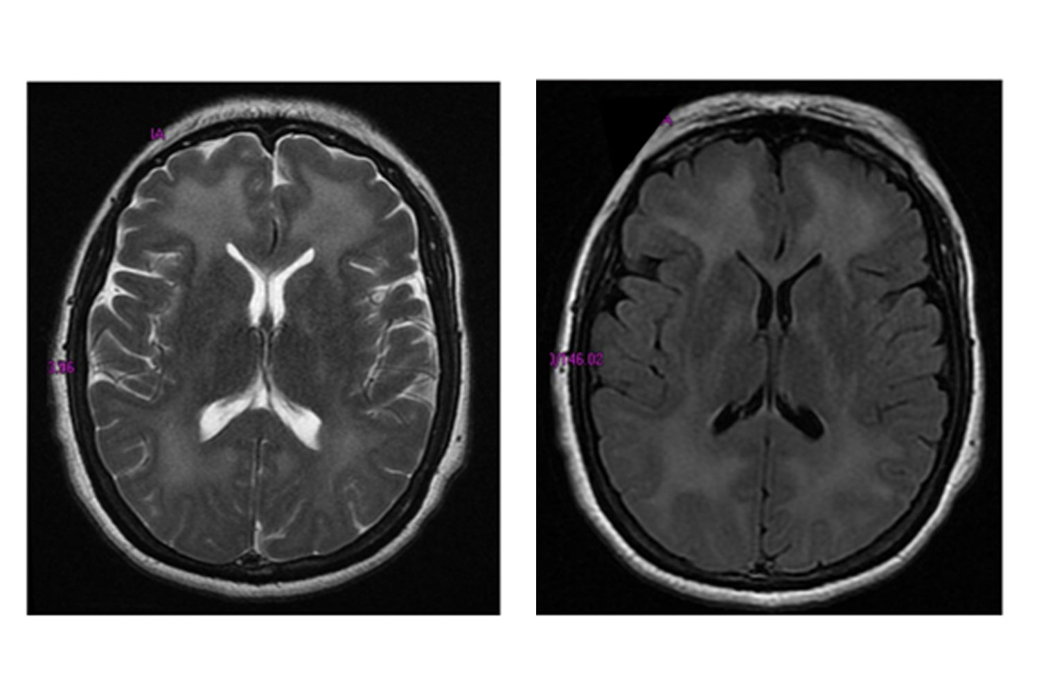
A 38 year old HIV-infected male (CD4 of 3 cells/μL) was treated for cryptococcal meningitis in Kampala, Uganda. The patient completed two weeks of standard amphotericin-based treatment for cryptococcosis without complications and was discharged. His only complaint at time of discharge was mild visual blurring. Combination antiretroviral therapy (ART) was initiated 5 weeks after cryptococcal diagnosis. Three months after starting ART, the patient presented to clinic complaining of headache, persistent visual blurring, vomiting, and a focal seizure. Brain CT with contrast at the time was normal (not shown), and the patient was started on anti-convulsant medication. A lumbar puncture was performed and CSF culture did not grow Cryptococcus neoformans (or any other pathogen). His symptoms continued to worsen over the next three months, and he was re-hospitalized 6 months from time of initial cryptococcal diagnosis. A lumbar puncture was repeated. The CSF remained sterile, VDRL and PCR testing for herpes simplex virus (HSV), varicella zoster virus (VZV), and cytomegalovirus (CMV) were negative. Post-gadolinium T2 sequences on brain MRI showed multiple irregular and patchy gyral enhanced lesions with marked effects noted in bilateral frontal, parietal, occipital, temporal lobes, and the basal ganglia (Figure 1a). Based on the timing of ART, clinical course, and imaging, a presumptive diagnosis of Immune Reconstitution Inflammatory Syndrome (IRIS) was given. The patient was started on prednisone (60 mg daily), and although the lesions persisted on follow-up MRI 4 months later (10 months after initial cryptococcal diagnosis; Figure 1b), the patient has experienced a slow, steady improvement in symptoms and remains in HIV care 28 months after his initial AIDS-defining diagnosis.
*Corresponding author: David R Boulware, Division of Infectious Disease and International Health, Department of Medicine, University of Minnesota, Minneapolis, MN, USA, E-Mail: boulw001@umn.edu
 Awards Nomination

