PHONE
+44-7482-878921
+44-7482-878921
2376-0249
Clinical Image - International Journal of Clinical & Medical Images (2017) Volume 4, Issue 10

Author(s): Madeeha Subhan* and Waleed Sadiq
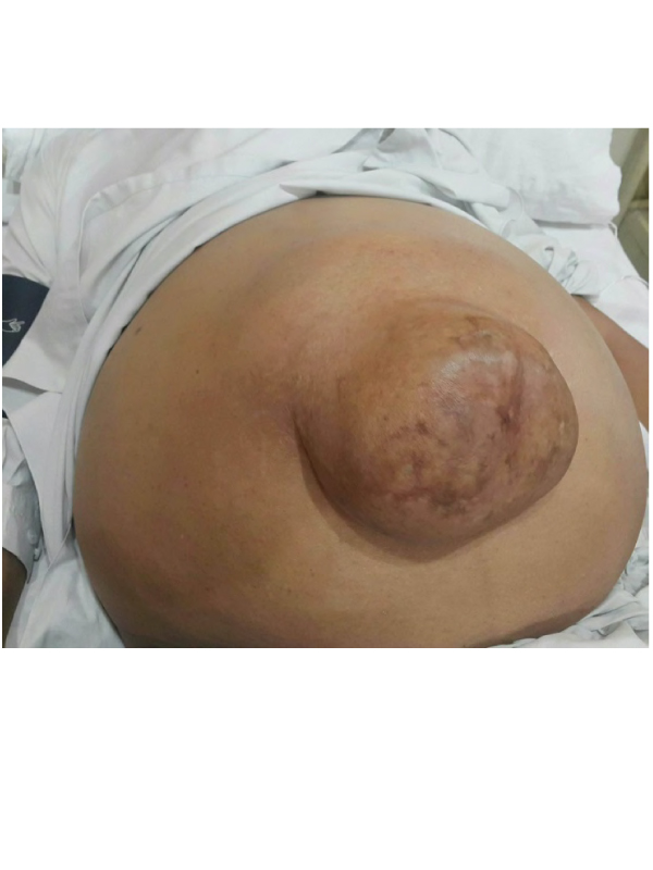
Clinical Image: A 58 year old male with supraumbilical reducible hernia, a year history of chronic liver disease presented to the outpatient department (Figure 1). He was diagnosed to be hepatitis C positive. On examination he had abdominal girth of 52 inches and it was reducible and cough impulse was positive. The hernia was 15 cm × 15 cm. His PCR-HCV RNA showed a viral load of 2192297 copies/ml. His ultrasound showed liver to be coarse granular echo rich parenchymal texture with slightly irregular margins and portal vein to be dilated (1.3 cm). Moderate abdominal ascites with defect in the linea alba with protruding gut loops and ascitic fluid in the hernia sac. The defect measures about 5 × 5.6 cm. His platelet count was decreased to 76 × 103/ul. Therapeutic paracentesis was done and mesh hernioplasty was done for hernia. Final Diagnosis: Paraumbilical hernia due to ascites caused by hepatitis C. Differential diagnosis: Malignancy, Tuberculosis, Lipoma.
 Awards Nomination