PHONE
+44-7482-878921
+44-7482-878921
2376-0249
Case Blog - International Journal of Clinical & Medical Images (2015) Volume 2, Issue 5

Author(s): Maria Fincati, Alexander KC Leung* and Benjamin Barankin
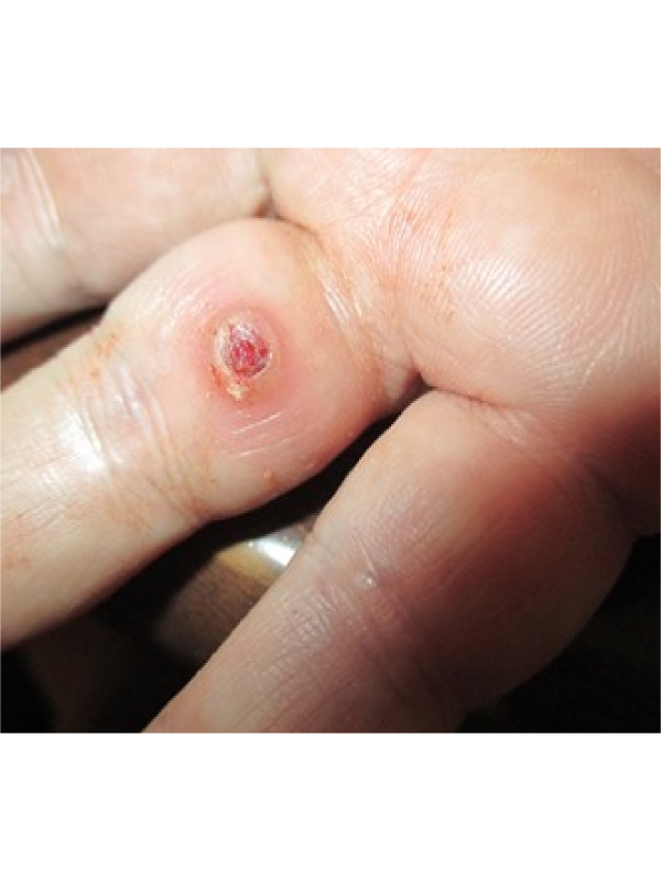
A 28 year-old woman presented with an erythematous, soft, dome shaped, red papule surrounded by a “collar” with a smooth friable surface on the right middle finger. The papule was asymptomatic, tended to bleed freely and had grown rapidly over the past three weeks. Pyogenic granuloma, also known as lobular capillary hemangioma, is a common, idiopathic, and acquired benign vascular tumor of skin or mucosa. Cutaneous lesions comprise approximately 0.5% of all skin nodules in children with a mean age of onset of 6.7 years. Adult incidence peaks in the third decade. Mucosal lesions termed granuloma gravidarum appear in 2 to 5% of pregnancies typically in the second and third trimester. Reactive response to trauma (e.g. ingrown nails), female sex hormones, and medications such as isotretinoin, may have a role to play in the pathogenesis. Clinically, pyogenic granuloma presents as a soft, dome-shaped papule/nodule or a sessile or pedunculated papule/nodule with a smooth, glistening, erosive, or friable surface. The color is usually bright red to dusky red initially. With time, the vascularity decreases and the lesion tends to become more collagenized and pink. Characteristically, the lesion is asymptomatic and painless. Due to its pronounced vascularity, pyogenic granuloma tends to bleed and ulcerate even with very minor trauma. Cutaneous lesions are commonly present located on the head and neck, trunk, and extremities, especially the fingers. Mucosal lesions are commonly oral and gingival. Nasal lesions are typically septal. Pyogenic granulomas rarely occur in the gastrointestinal tract, genital urinary tract, and the CNS.
Diagnosis is mainly clinical; consider referral to a dermatologist if diagnosis is in doubt and for treatment. Dermoscopy reveals a red homogeneous area and white scaly collarette. Although cutaneous pyogenic granuloma have no malignant potential, biopsy is warranted if the diagnosis is in doubt to rule out amelanocytic melanoma. Histopathology reveals a central feeder vessel surrounded by a proliferation of capillaries with areas of regression and extensive fibrosis. Pregnancy related lesions generally resolve spontaneously after childbirth, although many women still choose to have their lesions treated. Surgical excision affords a pathological specimen, has the lowest recurrence rate, and is the treatment of choice. Other treatment options include laser surgery, electrodessication, curettage, cryotherapy, topical silver nitrate, and topical imiquimod. Recurrences may occur largely attributed to inadequate primary destruction.
 Awards Nomination