2376-0249
Case Blog - International Journal of Clinical & Medical Images (2016) Volume 3, Issue 7

Author(s): Karthik S, Sekar B and Parthipan G
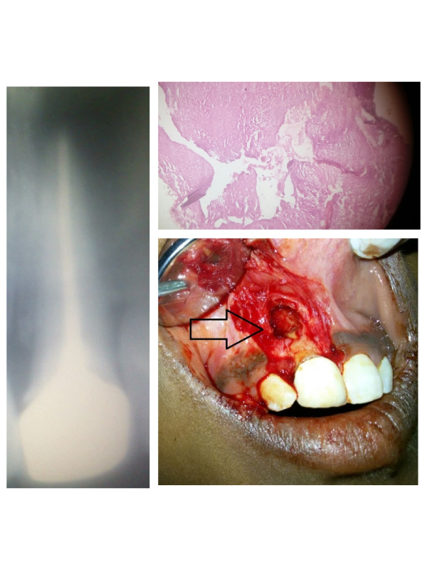
Clinical Presentation: A 25 year old female presented to the Department of Dentistry and Faciomaxillary Surgery complaining of discomfort in the region between labial surface of upper anterior teeth and inner mucosal layer of upper lip in relation to first quadrant. General health examination revealed that the patient was conscious, oriented, afebrile and vitals were stable. On intraoral clinical examination, a smooth lobulated slow enlarging spheroidal or ovoid mass was observed labially in relation to right maxillary central incisor tooth. Patient stated that the swelling has started as a pinpoint lesion before 6 months and progressed to the size of 2 cm × 3 cm × 1 cm as seen during clinical examination. The colour of the mass was of coral pink, smooth, symptomatic and the affected tooth was non-vital. Radiologically, round to oval radiolucency followed by well-defined cortical border (Figure 1). Clinical and radiological diagnosis claimed to be of radicular cyst based on its pathognomonic features which was later confirmed by histopathology. Histopathologically, stratified squamous epithelium, Rushton hyaline bodies, cholesterol cleft, fibrous capsule and scattered ciliated cells were seen (Figure 2). Conventional surgical enucleation was the preferred method of treatment (Figure 3). A mucoperiosteal flap over the cyst region raised followed by bony window created for access, careful separation of cyst fromits bony wall, removal of the entire cyst intact, smoothening of the bony cavity edges, controlled free bleeding, irrigation of the cavity to remove the remaining debris, replacement of the mucoperiosteal flap and sutures in place. Postoperative review was after 1 month with good prognosis thereby facilitating successful outcome. The radicular cyst is the most common cyst of the jaw. Etiology includes pulpal necrosis, dental caries and trauma. Source is linked to the Cell Rests of Malassez [1]. Radiographically, seen as a radiolucency sorrounded by radiopaque region in relation to the periapical region of the involved tooth. Commonly three phases are seen, initiation, formation and enlargement. Actinomyces for
 Awards Nomination

