PHONE
+44-7482-878921
+44-7482-878921
2376-0249
Case Blog - International Journal of Clinical & Medical Images (2015) Volume 2, Issue 4

Author(s): Ramachandra Reddy VJ*, Usha MK, Jayaranganath M and Manjunath CN
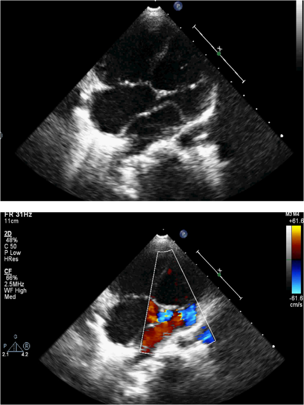
Triatrial heart is a rare congenital abnormality, reported in 0.4% of patients with congenital heart disease [1], and in less than 0.1% of clinically diagnosed cardiopathies [2]. First described by Church in 1868, the name “cor triatriatum” was given by Borst in 1905 [3]. It involves usually the left atrium (cor triatriatum sinister) and rarely the right atrium (cor triatriatum dexter). In the pediatric population may be associated with major congenital cardiac lesions such as tetralogy of Fallot, double outlet right ventricle, coarctation of the aorta, partial anomalous pulmonary venous connection, persistent left superior vena cava with unroofed coronary sinus, ventricular septal defect, atrioventricular septal defect, and common atrioventricular canal [4]. Clinical manifestations depend upon the size of the opening in the septum and the presence of associated congenital cardiac defects. We report a case of 5 years old girl who presented with history of exertional dyspnea ,fatigue and cyanosis. Echocardiogram done showed membrane attached laterally to the junction of the left upper pulmonic vein and left atrial appendage, dividing the left atrium into 2 chambers (Figure 1).
The proximal chamber receives blood from the pulmonary veins and the distal chamber contains the left atrial appendage and mitral valve with single fenestration communicating both chambers (Figure 2). Contrast echo was done in view of cyanosis with agitated saline showed early appearance of bubbles in left atrium before right atrium suggestive of unroofed coronary sinus. Left sided superior venacava (SVC) was also seen. Diagnosis of cor-triatrium sinistrum with unroofed coronary sinus and left SVC was made and surgery was advised. Diagnosis of cor triatriatum is frequently made with considerable delay due to rarity of the condition and presenting signs and symptoms that mimic other more common cardiac or pulmonary disorders. A high index of suspicion can prevent delayed diagnosis and unfavorable outcomes.
 Awards Nomination