PHONE
+44-7482-878921
+44-7482-878921
2376-0249
Clinical-Medical Image - International Journal of Clinical & Medical Images (2022) Volume 9, Issue 7

Author(s): Ana Sofia Alves*
Department of Radiology, University of Beira Interior, Covilhã, Portugal
Date of Submission: 28 June 2022, Manuscript No. ijcmi-22-67954; Editor assigned: 01 July 2022, Pre QC No. P-67954; Reviewed: 08 July 2022, QC No. Q-67954; Revised: 15 July 2022, Manuscript No. R-67954; Published: 22 July 2022, DOI: 10.4172/2376-0249.1000840
Citation: Alves AS. (2022) Seizures and Bilateral Basal Ganglia Calcifications Following Thyroidectomy. Int J Clin Med Imaging 9:840.
Copyright: © 2022 Alves AS. This is an open-access article distributed under the terms of the Creative Commons Attribution License, which permits unrestricted use, distribution, and reproduction in any medium, provided the original author and source are credited.
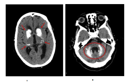
A 72-year-old woman with a past medical history of hypertension, dyslipidaemia, atrial fibrillation, heart failure and epilepsy for the last 3 years and partial thyroidectomy as of 20 years ago. She presented herself at the emergency room with a 9-month history of seizures and psychosis and was admitted for investigation. The physical examination showed neglect. Laboratory testing revealed hypocalcaemia 7.2 mg/dl (normal value, <8.8) with hyperphosphatemia 6.9 mg/dl (normal value, <4.5) and hypoparathyroidism 3.7 pg/ml (normal value, <10). Vitamin D, magnesium, albumin, and thyroid functioning testing came back normal. The brain computer tomography (CT) revealed bilateral basal ganglia calcifications (Figure 1A and 1B). Fahr’s syndrome was diagnosed.
The diagnosed of Fahr’s syndrome was assumed considering her past medical history, the clinical presentation, physical examination findings, and subsequent investigation exams. The aetiology assumed was secondary to hypoparathyroidism related to her prior partial thyroidectomy [1]. She started treatment with calcium and calcitriol with improvement.
By using this particular case, authors aim at alerting that the presence of neuro-psychiatric symptoms and basal ganglia calcifications, Fahr’s syndrome should be considered even if rare, especially after thyroidectomy [2]. Sometimes, these symptoms attributed to dementia and some patients can be undiagnosed and not receive the correct treatment.
Also, it is important to note that there are secondary forms, such as this one, and usually these are related to abnormal calcium/phosphorus (Ca/P) homeostasis. In this case, it was due to the involuntary removal of the parathyroid glands during thyroidectomy.
The treatment should direct to the relief of symptoms and pointed to the under-lying cause. In the presence of abnormal Ca/P or parathyroid disorder, the treatment involves calcium and calcitriol, also evident in the advanced stage of this disease. Prompt treatment can prevent brain calcifications and neurophysiological disorders [3].
Bilateral basal ganglia; Thyroidectomy
Google Scholar, Crossref, Indexed at
Google Scholar, Crossref, Indexed at
 Awards Nomination