2376-0249
Clinical Image - International Journal of Clinical & Medical Images (2014) Volume 1, Issue 7

Author(s): Int J Clin Med Imaging 2014
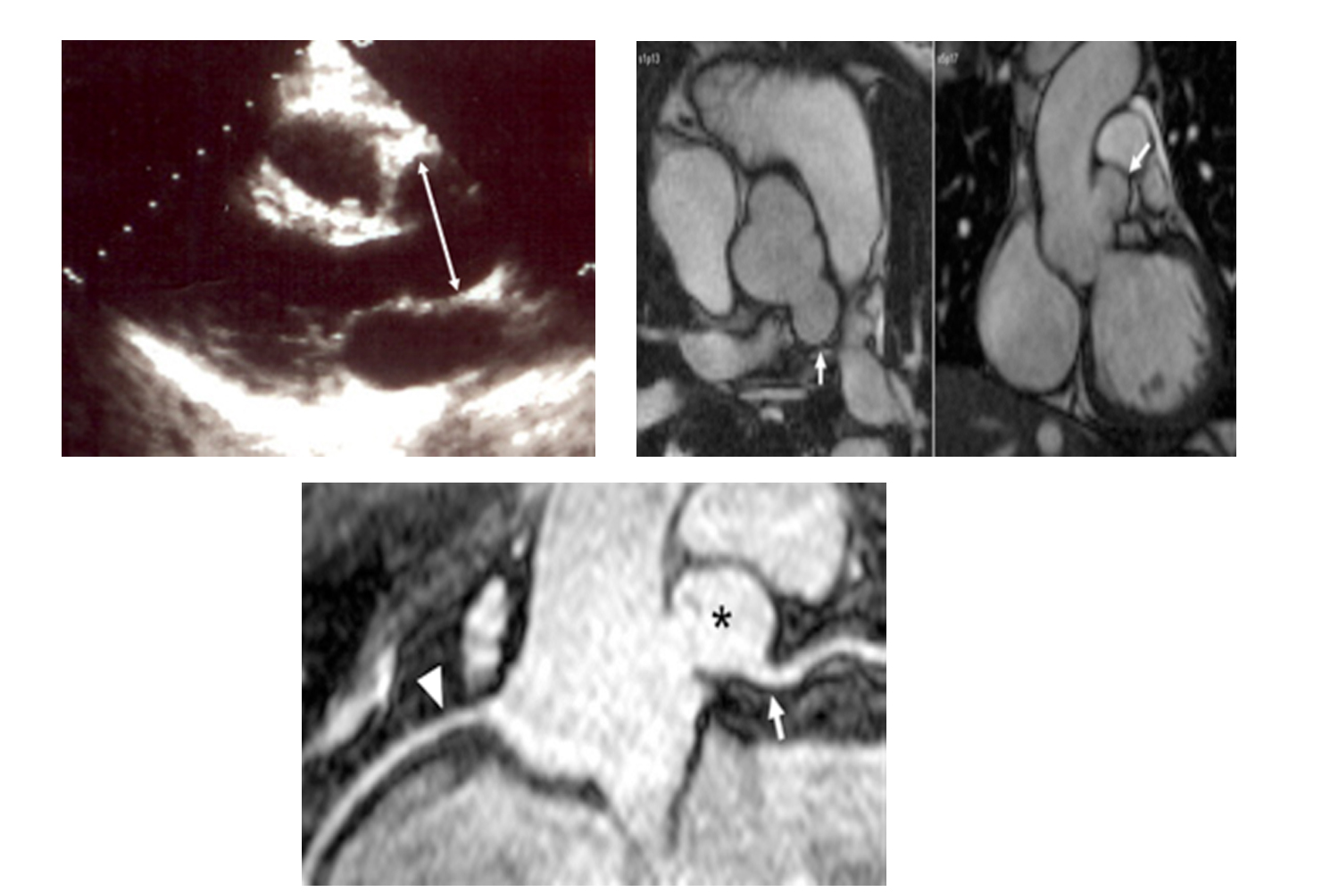
A 28 year-old asymptomatic female with no significant medical or family history received a transthoracic echocardiogram for “practice” while accompanying her mother to a cardiologist’s office. She was found to have a dilated aortic root (Figure 1) measuring 4.1 cm at the Sinuses of Valsalva, with normal left ventricular function and no aortic regurgitation. Cardiac Magnetic Resonance Steady State Free Precession cine imaging revealed an aneurysm of the left sinus of Valsalva measuring 2.0×1.9 cm (Figure 2), but the location of the coronary ostium in relation to the aneurysm was difficult to determine. Free breathing 3D navigator guided technique was performed for the delineation of whole heart coronary anatomy without contrast at high spatial resolution, with a voxel size of 0.55 x 0.55 x 0.80 mm3. A curved linear reformat (Figure 3) revealed that the left main coronary (arrow) originated from the aneurysm wall (asterisk). The normal right coronary artery (arrowhead) is also shown. Surgical patch repair would have necessitated left main coronary excision and re-implantation, so the decision was made to forego surgery and follow the patient with periodic observation. Patients diagnosed with sinus of Valsalva aneurysms commonly present in the context of symptomatic aneurysm rupture into an adjacent cardiac chamber [1].
 Awards Nomination

