PHONE
+44-7482-878921
+44-7482-878921
2376-0249
Case Blog - International Journal of Clinical & Medical Images (2014) Volume 1, Issue 4

Author(s): Int J Clin Med Imaging 2014
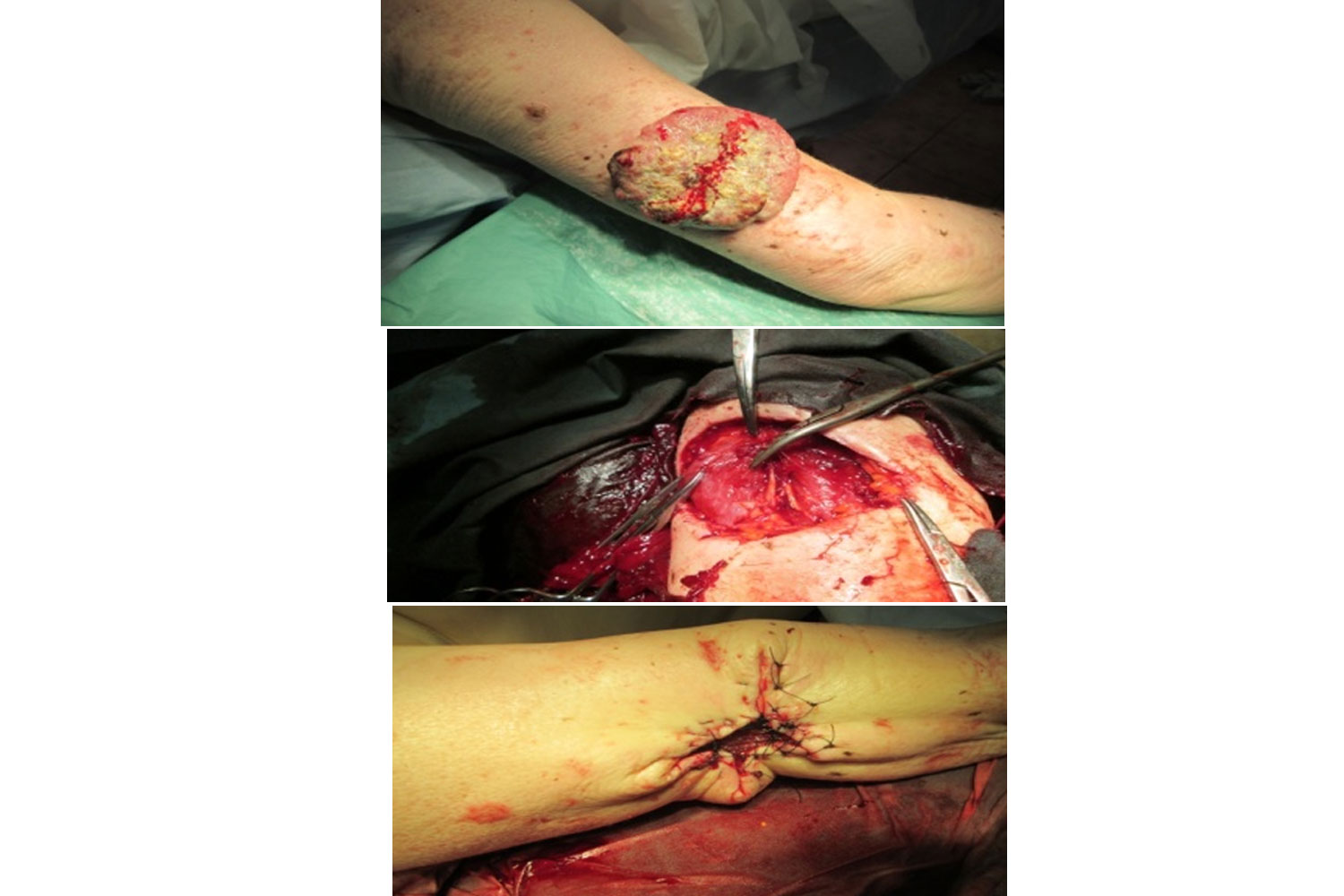
A 30-year old albino woman was referred by a dermatologist for excision of a large ulcerated and fungating squamous cell carcinoma (6 x 5cm) on the outer aspect of the right arm. (Figure 1) There was neither palpable axillary lymphadenopathy nor evidence of distant metastases and, she was otherwise clinically well. Successful removal necessitated the excision of a sufficiently wide margin of surrounding tissue (1cm) with the primary growth. The tumour was seen to invade the deep fascia of the underlying muscle and careful fasciectomy with sparing of the musculocutaneous nerve was performed. (Figure 2) The resulting large defect in the tight compartment of the arm made closure difficult. Skin grafting was not done in the uncertainty of adequate clearance of the malignant lesion and certainly not directly over muscle. However, to facilitate healing, careful undermining of the adjacent tissues permitted partial closure of the defect. (Figure 3) The wound healed successfully within 4 weeks and there was no evidence of local recurrence 5 months later. Albinism is an inherited autosomal recessive disorder characterized by lack of skin pigmentation. It appears to be the most important risk factor in the development of squamous cell carcinoma through the malignant transformation of active keratosis 9 [1,2]. It is also the commonest cause of death in young African albinos [2]. Treatment entails locoregional control by surgical excision along with block dissection of involved regional glands [3]. Radiotherapy alone may provide palliation when gland dissection is impracticable.
 Awards Nomination