2376-0249
Case Blog - International Journal of Clinical & Medical Images (2016) Volume 3, Issue 1

Author(s): Hakan Demirtas*, Ahmet Orhan �elik, Ayşe Umul and Mustafa Kara
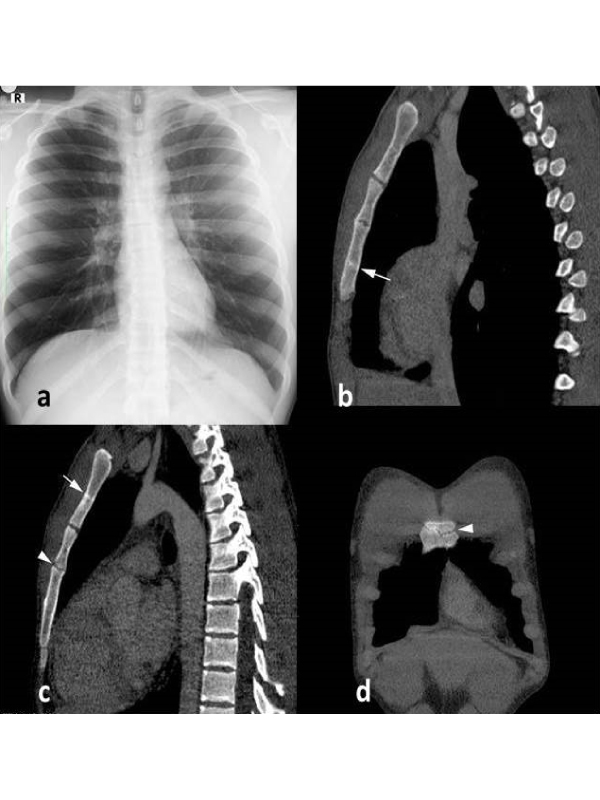
Clinical Presentation A 22 year old male patient, involved with wrestling, admitted to our centre due to the complaints of anterior chest wall pain and difficulty in breathing. No apparent finding was found during physical examination of the patient, except for tenderness in the sternal region. No specific findings were detected in the patient’s routine blood test, and results of the patient’s respiratory tests were normal. Chest radiogram was normal (Figure 1A). Stress fractures (Figure 1B and 1C) and an acute fracture line (Figure 1D) were observed in the sternal region in MDCT performed for the thorax. Stress fractures that usually occur in athletes who are exposed to overloading in unfavourable grounds are observed particularly in below knee structures depending on recurrent micro-traumas. In contrast to acute fractures caused by a single strong loading, stress fracture is caused by strong non-recurrent pressures [1]. Stress fractures may be suspected in symptomatic patients without any actual fracture line. But, actual stress fractures may occur if loading continues. This type of fracture is relatively rare among athletic injuries (1-7%) [1] and observed particularly in military recruits, runners, those interested in jumping sports and involved with repetitive loading activities. Stress fracture in the sternal region is observed at the rate of 0.5% in all of sternal fractures and is very rare [2]. This type of fracture is defined in gymnasts and military recruits in the literature, and only a single case has been reported in those involved with wrestling to the extent our knowledge [3]. Diagnosis of this type of fracture is established by clinical, physical examination and imaging methods. Imaging studies including radiographs, computed tomography (CT) scans, MRI, and bone scintigraphy can be helpful in diagnosis.
 Awards Nomination

