PHONE
+44-7482-878921
+44-7482-878921
2376-0249
Clinical-Medical Image - International Journal of Clinical & Medical Images (2016) Volume 3, Issue 9

Author(s): Marco Di Serafino*, Valeria Coppola, Francesco Lisanti, Rosario Rocca and Enrico Scarano
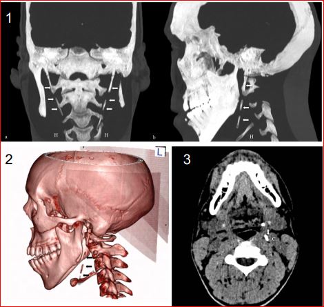
A 30 year-old man presented at our department complaining of two years of bilateral neck pain, greater during swallowing, chewing and stretching head. There was no history of head trauma or surgery. He was treated unsuccessfully for a trigeminal neuralgia. Clinical examination of oral cavity and oropharynx revealed no abnormalities or cervical lymph nodes, but an exacerbation of pain during palpation of tonsillar fossae. Non-contrast computed tomography (CT) scan showed bilaterally elongated styloid processes with their lower tips at the level of hyoid bone anteriorly, along the inferior margin of the base of tongue measuring 9 cm (Figures 1 and 2); they were close to the carotid-jugular complex (Figure 3). We made a diagnosis of Eagle’s Syndrome. The complete removal of elongated stylohyoid ligaments brought to clinical resolution. It is very important for all clinicians to include Eagle’s syndrome in the differential diagnosis when treating patients with atypical pain in the head and neck regions.Eagle syndrome is a rare condition where elongated temporal styloid processes (> 3 cm), or calcified stylohyoid ligaments, are in conflict with the adjacent anatomical structures giving rise to a complex range of variable symptoms including otalgia, dysphagia, foreign body sensation in throat, pain along carotid artery distribution and others [1,2]. Its diagnosis is clinical and radiological both with orthopantomogram and conventional radiographs both with Computed Tomography (CT). The latter is recommended because it shows better the detailed anatomy thanks to its multi-parametric and 3D-volume rendering reconstructions which support the management, planning and tailoring of the surgical approach [1,2]. The treatment plan includes both medical and surgical options.
 Awards Nomination