PHONE
+44-7482-878921
+44-7482-878921
2376-0249
Clinical-Medical Image - International Journal of Clinical & Medical Images (2021) Volume 8, Issue 12

Author(s): Drissi Abdel-ilah, Merbouh Sahar and Sara Habibchorfa
Clinical-medical Image
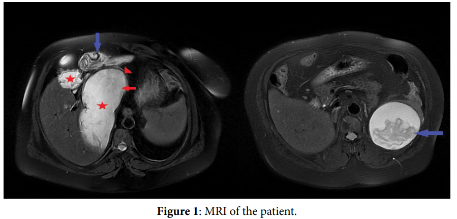
A 65-year-old man operated for hydrated cyst with cholecystectomy 8 years ago, control ultrasound found a multiple hepatosplenic cystic formations, an abdominal MRI was requested which revealed a multiple hepatic and spleno-peritoneal hydrated cysts with a floating membrane inside realizing the appearance of the snake sign (Figure 1). The snake sign is a pathognomonic radiological sign of hydrated cysts which are structures due to Echinococcus Infections; these structures represent the laminated membranes of the end cyst detached from the priciest, resulting in intracystic floating membranes that mimic the appearance of a serpent or a spin whirl sign. The typical hydrated cyst is made up of three layers: an outer fibrous capsule (priciest), made up of cells that form a rigid protective zone, a laminated middle a cellular membrane (exorcist) that allows the passage of nutrients and can be easily ruptured, predisposing to infection and an inner germ layer (end cyst), where the solaces are located (the larval stage of the parasite). Detachment of the end cyst from the priciest may be secondary to several causes including the decreased intracystic pressure, degeneration, host response, trauma or response to medical therapy, and percutaneous drainage. X-rays can detect the “snake sign” in hydrated lung disease; however “serpentine membranes” in a hydrated cyst are classically described on abdominal ultrasound, computed tomography or MRI in particular the non-fat-saturated FIESTA section which show a lesion with hypo intense wall and intracystic hyper intense germinate membrane and floats inside, representing the ‘‘serpent sign’’.
Keywords: The snake sign, Hydrated cysts
Declaration of conflicting interests
The authors declared no potential conflicts of interest with respect to the research, authorship, and/or publication of this article.
 Awards Nomination