PHONE
+44-7482-878921
+44-7482-878921
2376-0249
Clinical Image - International Journal of Clinical & Medical Images (2014) Volume 1, Issue 3

Author(s): Ahmad Al-Awwad, Linda A. Hershey
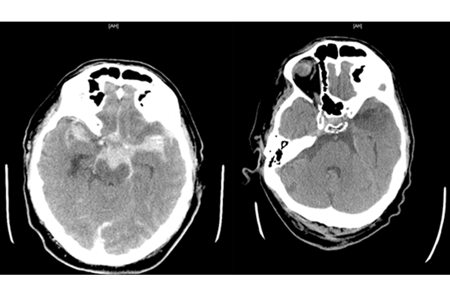
A 73 year old male horse trainer was admitted to the emergency department for acute traumatic subarachnoid hemorrhage after he was thrown from an untrained horse and fell on his head. The figure on the left is a CT that shows evidence of an acute subarachnoid hemorrhage. The patient was taking warfarin at the time of his fall, because he had an artificial aortic valve (INR=2.5). He was initially alert and oriented, but he quickly became confused, disoriented and drowsy. The cerebral angiogram on admission showed no signs of aneurysm or arterio-venous malformation. He had a complicated clinical course and developed hydrocephalus. Three weeks later (see figure on the right), the CT showed evidence for a left temporal lobe contusion. The patient was still disoriented to time and place, but he could follow 1-step commands. His wife was counselled at discharge about how he should neither drive, nor ride a horse again. When the patient was seen 10 months later, he was still was signitificantly cognitively impaired (MMSE=14/30).
blog
blog
blog
blog
blog
blog
blog
blog
blog
blog
blog
blog
blog
blog
blog
blog
blog
blog
blog
blog
blog
blog
blog
blog
blog
blog
blog
blog
blog
blog
blog
blog
blog
blog
blog
blog
blog
blog
blog
blog
blog
blog
blog
blog
blog
blog
blog
blog
blog
blog
Corresponding author
Ahmad Al-Awwad and Linda A. Hershey
University of Oklahoma Health Sciences Center
USA
 Awards Nomination