PHONE
+44-7482-878921
+44-7482-878921
2376-0249
Clinical Image - International Journal of Clinical & Medical Images (2014) Volume 1, Issue 4

Author(s): Int J Clin Med Imaging 2014
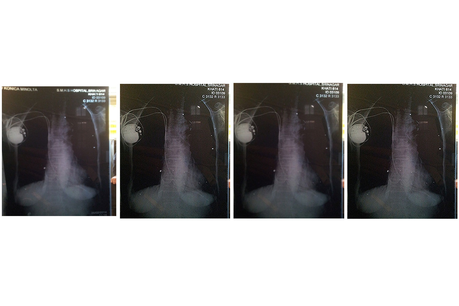
A 75 years old female hypertension, Type 2 diabetes mellitus, on treatment. Patient was put on permanent pacemaker for symptomatic complete heart block in 2006.she reported again to causality with complete heart block with history of intermittent presyncope for 1 year. Evaluated ECG showed complete heart block. Chest x-ray showed pacemaker lead in the abdominal cavity piercing right ventricular myocardium and diaphragm of chest and lied in the abdominal cavity adjacent to spleen. Echocardiogram showed the pacemaker lead piercing the right ventricular myocardium and diaphragm of the chest into the abdominal cavity, there was no hemocardium or pericardial effusion. The patient was taken to cardiac cath lab and fresh pacemaker lead was implanted and the previous lead kept in situ.
 Awards Nomination