2376-0249
Case Blog - International Journal of Clinical & Medical Images (2016) Volume 3, Issue 9

Author(s): R S Sisodiya, Simmi K Ratan and Parveen Kumar
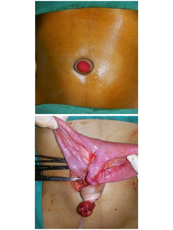
Case Presentation: An 8 year old male child belong to poor socioeconomic background came to pediatric surgical unit with complain of presence of mucoid discharging red fleshy mass in umbilicus since birth with occasional discharge of fecal matter(once in 3 to 4 month) from it. There was no history of vomiting, abdominal distension or any urinary problem. Child had previously consulted some medical practioner who mistook it as granuloma and advices reassurance and some local ointment application. As parent of child had financial constrain and further child have no serious problem, parent ignored his ailment and sought tertiary level medical care late. Examination revealed firm red mass in umbilicus, periumbilical skin was normal (Figure 1).
Keeping vitellointestinal duct (VID) remnant as most probable diagnosis Trans umbilical exploration was performed, there was patent vitellointestinal duct communicating with this polyp. Resection with primary anastomosis of ileum was done (Figure 2), post-operative recovery was uneventful, and biopsy was consistent with VID. Umbilical granuloma represents persistence of inflammatory reaction with granulation tissue formation in umbilical tissue that has yet to epithelialize. Generally they are small, moist, velvety masss [1]. While Patent VID represent rare form of omphalomesenteric duct remnant that require surgical treatment, the umbilical part of this duct looks like umbilical polyp (red firm mass) [2,3]. Commonly umbilical granuloma respond to cauterisation, rarely they require surgical removal in case they does not respond to cauterisation alternative diagnosis (VID remnant, urachal remnant) must be kept and accordingly managed [1].
Learning Points:
• Care full history and examination is of paramount importance in any umbilical mass assessment.
• Non response of umbilical granuloma to cauterisation must prompt physician to think alternate diagnosis.
• Patent VID need surgical intervention as delays in diagnosis put patient at risk of developing serious complication (volvulus).
 Awards Nomination

