PHONE
+44-7482-878921
+44-7482-878921
2376-0249
Clinical Image - International Journal of Clinical & Medical Images (2021) Volume 8, Issue 12

Author(s): Jairo Renteria Roa
Clinical Image
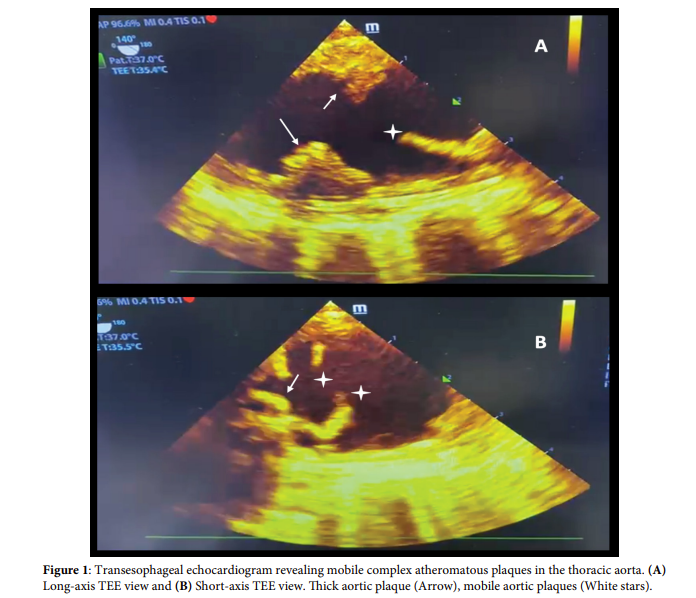
Aortic atherosclerotic atheromas usually compromise the descending aorta [1]. Complex or unstable aortic plaques are pedunculated, mobile, ulcerated, or thick (>4 mm) [2]; compared to flat plaques, the first is associated with a higher risk of embolic events [3]. Stroke, transient ischemic attack, and cardiac infarction are the commonest manifestations when the proximal aorta is involved [4]. A73-year-old woman with a history of hypertension, diabetes mellitus, dyslipidemia and heavy smoking, with documented three-vessel coronary artery disease, presenting as a non-ST acute myocardial infarction. A transesophageal echocardiogram (TEE) revealed a normal ejection fraction, extensive atherosclerosis of the aorta and two pedunculated plaques in the ascending thoracic aorta (Figure 1). Off-pump coronary artery bypass grafting was considered, but the patient was reluctant to the intervention so aspirin, clopidogrel, and high dose atorvastatin were started.
Pedunculated aortic plaques pathogenesis is associated with rupture of soft plaques, endothelial injury and organized thrombus formation [2]. Studies report up to a four-fold increased risk of embolic events with a relative-risk of 3.8 (95% CI 1.8-7.8) in the presence of grade 4 or 5 aortic atheromas [1]. TEE allows diagnosis and embolic risk stratification; computed tomography helps in planning surgical intervention [4]. Lipidlowering and antiplatelet medications are the cornerstone of treatment; smoking cessation, glycemic and hypertension control are mandatory [4]. Surgical procedures are controversial [4], patient eligibility is based on the risk of embolism and recurrence, because mortality associated with complex aortic plaque may be as high as 20% within three years without intervention [1] but immediate embolism to mesenteric, cranial o extremities circulation carries a high risk of death and morbidity [4]. The authors highlight this severe manifestation of systemic atherosclerosis.
Keywords: Mobile plaques; Thoracic aorta
Declaration of Interests The authors declare that they have no competing interests.
References
 Awards Nomination