PHONE
+44-7482-878921
+44-7482-878921
2376-0249
Medical Image - International Journal of Clinical & Medical Images (2016) Volume 3, Issue 3

Author(s): Massimo Bolognesi
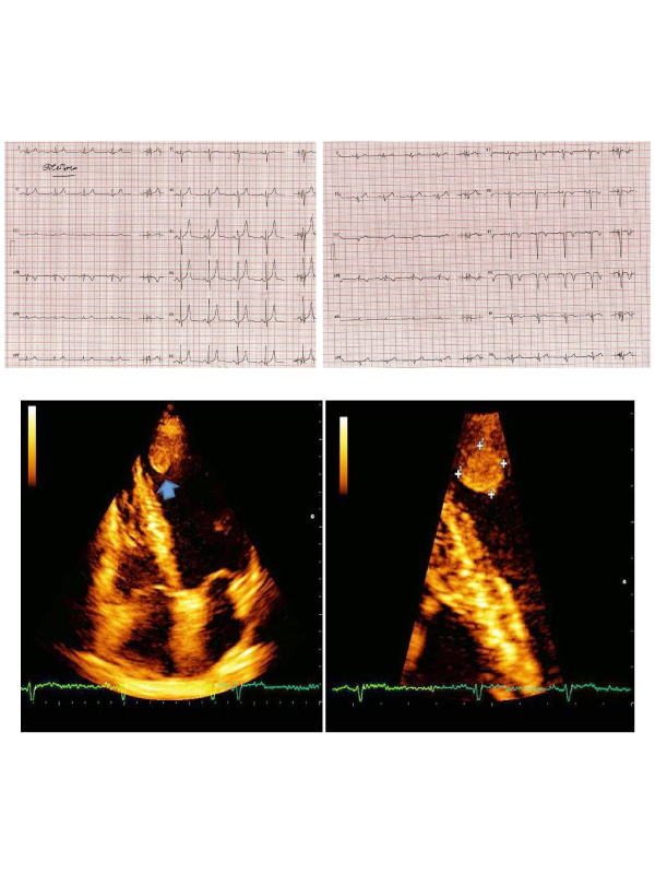
Figure 1: Old ECG.
Figure 2: Last ECG.
Figure 3: Focused 2D TT echocardiogram.
Herewith the author describes the history of a healthy 60-year-old amateur rider who came to our sports cardiology medicine center for sports pre-participation screening. His family history was unremarkable and physical examination was normal. He claimed he felt fit and that he was taking regular physical activity by cycling. His old ECG was normal (Figure 1) while the last ECG (Figure 2) was abnormal for findings of recent anterior myocardial infarction. A subsequent focused 2D TT echocardiogram through classical and magnified apical 4 chamber view showed left ventricular apical akinesis with small apical thrombus (Figure 3 - see arrow) protruding into the left ventricular cavity and demonstrates mobility in real-time imaging. The athlete was admitted to cath lab for revascularization procedure and anticoagulation therapy was started. The risk of cardioembolic stroke was very high.
 Awards Nomination