2376-0249
Clinical Image - International Journal of Clinical & Medical Images (2017) Volume 4, Issue 6

Author(s): Mayo RC
Introduction: Implanted medical devices are increasingly common, and it is important for radiologists to recognize the normal appearance of these devices in order to inspire confidence among patients and referring physicians. The goal of this article is to teach the appearance of wireless cardiac monitoring devices and briefly review their function.
Keywords: Wireless; Monitor; Breast; Cardiac device
Discussion: Heart disease is the number one cause of death in the United States [1]. Monitoring for cardiac arrhythmias is important since they may cause serious adverse events including stroke, pulmonary embolism, and even to sudden death [2]. Rapid detection is crucial for diagnosis and appropriate treatment. Traditional monitoring devices, like the Holter monitor, are cumbersome, expensive, and often ineffective [3]. The devices depicted in these are wireless cardiac monitors. They are able to wirelessly detect and record cardiac activity.
Some models contain integrated cellular technology that can transmit diagnostic data from the device to a physician’s office. They can even be programmed to send an alert to a smart phone [4,5]. They may be as small as one-third the size of an AAA battery and are designed to function for up to 3 years. Some models are safe to use in an MRI magnet [5,6]. Breast cancer is the second leading cause of cancer deaths in women [7]. There is a powerful overlapping risk factor that both breast cancer and cardiac disease has in common - age. Therefore the likelihood of seeing cardiac devices in this patient population will increase as biomedical technology advances. In fact many innovative companies are beginning to leverage technology to impact patient care [8]. As this trend progresses, the transition from population based care to individualized medicine tailored to the specific patient needs will continue [4]. The benefit of wireless cardiac monitors is that they allow both improved arrhythmia diagnosis and create the potential for individualized anticoagulation dosing rather than standard one-size-fits-all treatment. This approach to individualized therapy is undergoing evaluation currently in randomized trials [9].
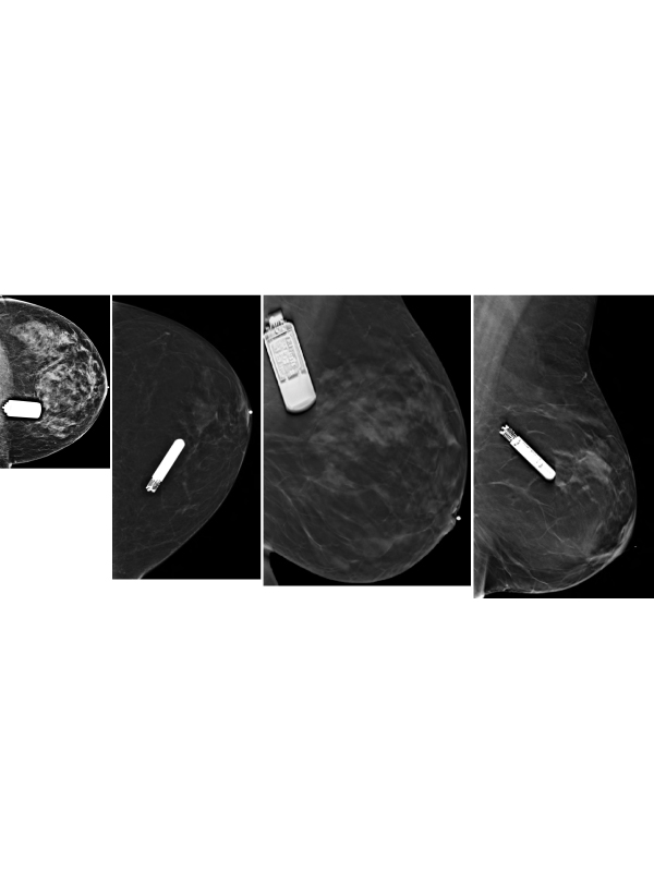
These devices are implanted in the outpatient setting. Under sterile technique a tiny incision is made in the left lower inner quadrant of the breast. Then a specialized insertion tool is used to position the device within the breast. Ideally the device is positioned 45o to the sternum at the 4th intercostal space 2 cm laterally to the sternum. As with any foreign device implantation, possible complications include device rejection, migration, infection, and hematoma formation [10]. The characteristic imaging appearance of these devices allows for confident identification. The first clue is its location in the left medial breast which allows close proximity to the heart for improved monitoring ability. The antenna component is identifiable at one end of the device and should be directed toward the chest wall posteriorly (Figures 1 and 2). Note that, tomosynthesis images (3D mammogram) shows the presence of some of the internal components, including a battery and circuitry (Figures 3 and 4). Unlike more traditional cardiac devices, there are no wires extending from this monitor. These findings are in contrast to older cardiac devices such as pacemakers and automated implantable cardiac defibrillators which also require a battery pack and wires leading to the heart.
 Awards Nomination

